刘玉霞 于振海
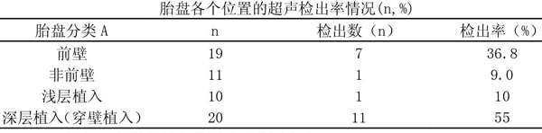
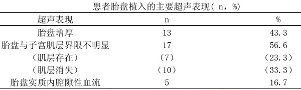
【摘? 要】目的:探討胎盘植入的超声图像特征及诊断价值,分析漏诊原因,以提高胎盘植入的产前超声诊断率。方法:对 30例产后确诊为胎盘植入的声像图进行回顾性分析。结果:1、产前彩色多普勒超声对前壁肌层胎盘植入的诊断率及检出率明显高于非前壁肌层胎盘植入。2、胎盘与子宫肌层界限欠清晰在胎盘植入的表现(胎盘增厚、胎盘与子宫肌层界限不清晰以及胎盘实质内腔隙性血流情况)中占主要部分。3、中央型前置胎盘的胎盘植入在前置胎盘中的检出率较高。结论:胎盘植入的超声声像图特征复杂多样,仍需进一步提高临床的诊断率及检出率; 前壁肌层的胎盘植入以及中央型前置胎盘有利于超声的诊断及检出。
【关键词】胎盘植入;超声
【中图分类号】R714.46????? 【文献标识码】A????? 【文章编号】1672-3783(2019)03-0021-02
【Abstract】Objective:To investigate the ultrasonographic features and diagnostic value of placenta implantation, and to analyze the causes of missed diagnosis, so as to improve the prenatal ultrasonographic diagnostic rate of placenta implantation..Methods: the sonographic findings of 30 cases of postpartum placenta accreta were retrospectively analyzed.Results: The diagnosis rate and detection rate of placenta implantation in anterior muscle layer by prenatal color Doppler ultrasound were significantly higher than that of non-anterior muscle layer placenta implantation. 2, the placenta and uterine muscle layer boundary is not clear in the placenta implantation performance(placenta thickening, placenta and uterine muscle layer boundary is not clear, and placenta parenchymal lacunal blood flow situation) is the main part. The rate of placenta implantation in the central placenta is higher than that in the placenta.Conclusion:The ultrasonic imaging features of placental implantation are complex and varied, and the diagnostic and detection rates still need to be further improved. The placenta implantation of the anterior muscle layer and the central anterior placenta are conducive to the diagnosis and detection of ultrasound.
【Keyword】placenta accreta; ultrasonic
引言
近年来,随着二胎政策的放开,剖宫产率呈现逐年上升的趋势,胎盘植入的发生率也大幅上升。胎盘植入对孕产妇的生命安全及健康情况产生严重威胁。超声应该注重对胎盘植入孕产妇的早期诊断,然而胎盘植入通常缺少典型的超声声像图特征诊断表现,为最大可能的提高胎盘植入的医学影像学诊断率及检出率,本研究采用三维超声和二维超声对妊娠期胎盘植入孕产妇进行检查,统计以及分析二维超声与病理结果的诊断符合率,分析以及总结胎盘植入的声像图特征。现将临床资料报道如下。
1 资料与方法
1.1临床资料
产后经手术证实为胎盘植入患者30例,年龄22~41岁,孕周14~41周,孕次1~4次,初产妇6例,经产妇24例;合并瘢痕子宫21例,前置胎盘11例,重度子痫1例。产前曾在我院进行B超检查1次14例,2次8例,3次以上8例。
1.2检查方法
使用VOLUSONGE8、VOLUSONGE10、VOLUSON730、IU22彩色多普勒超声诊断仪,探头频率为 5 ~ 1M H z(腹部), 8~ 4MHz(经阴道)。孕产妇保持平卧位,通过进行二维常规腹部超声扫描,必要时进行阴道超声,并辅以彩色多普勒和(或)三维超声作进一步检查。通过定量以及定性分析胎盘的位置、厚度以及成熟度,仔细观察胎盘与子宫肌层的边界以及有无异常回声的存在(包括后间隙低回声区)、子宫浆膜层和膀胱壁高回声区以及完整性,子宫肌层中液体暗区,仔细观察子宫肌层和实质内血流改变和血流量。
- 先锋戏剧在文化产业上的地位与发展趋势分析
- 地域文化发展与当代文学作品的关系
- 论水的意象对生命本真的诠释
- 《辽史乐志》中的“四旦”之争论
- 从《梦游天姥吟留别》看唐道教文化对李白的影响
- 弱势群体残疾人职业教育及就业研究
- 质谱分析技术在纺织品检测中的应用
- 构建高校教师党支部书记“双带头人”培育工程长效机制的思考
- 浅谈满族民居建筑装饰的特点
- 基于空间句法的博物馆建筑内部空间认知分析
- 优势、效果和问题:虚拟现实(VR)技术与广告传播研究
- 艺术高职院校贫困生资助工作的现状及发展研究
- 谈导引标识系统建设
- 浅析“经营位置”与阿尔茨海默症宜居空间的构建
- 高校大学生事务管理服务体系的建构探索与实践
- 新时期高校学生党员组织生活创新研究
- 词四首
- 相亲
- 又被蒺藜扎了
- 为了自己,突破自己
- 为众人抱薪者不可使其厄于风雪
- 每个人都应该更好的“努力”
- 地域文化与群众文化活动的融合
- 浅析农村群众文化工作中存在的问题及对策建议
- 基层群众文化活动组织及辅导研究
- transferals
- transferbook
- transfer book
- transfer deed
- transferdeed
- transferee
- transferincome
- transfer income
- transfer of training
- transferoftraining
- transferofundertaking
- transfer of undertaking
- transferor
- transfer payment
- transferpayment
- transferprice
- transfer price
- transfer pricing
- transferred
- transfer register
- transferregister
- transferrers
- transferring
- transfers
- transfer²
- 叙会
- 叙写记载
- 叙利亚
- 叙别
- 叙功
- 叙効
- 叙家常
- 叙常
- 叙录
- 叙悲
- 叙情
- 叙意
- 叙才
- 叙文
- 叙断
- 叙旧
- 叙晤
- 叙梦
- 叙爵
- 叙用
- 叙礼
- 叙离
- 叙称
- 叙绩
- 叙致