张凯旋 陈广新 邱收


摘 ?要: 探讨椎动脉阻断前后基底动脉瘤的血流动力学变化。采集基底动脉瘤患者颅内CTA影像DICOM格式数据,应用MIMICS 21.0软件三维重建椎动脉模型,应用3-matic软件对初步获得的模型进行修复,并使用Ansys ICEM软件对模型进行离散化网格划分,最后通过Ansys fluent软件对动脉瘤有限元模型进行数值模拟运算,获得颅内正常动脉与术前、术后动脉瘤部位血流速度、壁切应力、壁压力的分布特征,比较分析正常、术前、术后三种模型之间的血流动力学参数差异。椎动脉阻断前后基底动脉瘤的血流动力学参数存在显著差异,三种模型的血流速度、壁切应力、壁压力分别两两之间差异具有统计学意义(P<0.05)。通过椎动脉阻断的方式,基底动脉瘤的血流动力学发生显著的改变:血流速度下降、壁切应力升高、壁压力下降。
关键词: 颅内动脉瘤;血流动力学;数值模拟;计算流体力学
中图分类号: TP319 ? ?文献标识码: A ? ?DOI:10.3969/j.issn.1003-6970.2019.06.021
本文著录格式:张凯旋,陈广新,邱收,等. 椎动脉阻断术前后基底动脉瘤的血流动力学数值模拟分析[J]. 软件,2019,40(6):96100
【Abstract】: To investigate the hemodynamic changes of basilar aneurysms before and after vertebral artery occlusion. Using Mimics21.0 import case of patients with intracranial aneurysms CTA images DICOM format data, obtain the intracranial aneurysm model, will import the model 3-matic model to build further, Ansys workbench to build a good model for grid division, numerical simulation calculation, the gain of intracranial aneurysm of blood flow velocity, wall pressure and wall shear stress (WSS) values, compare and analyze the hemodynamic parameters of the differences between the three models. There were differences in hemodynamic parameters of basilar aneurysm before and after vertebral artery occlusion, and the differences in blood flow velocity, WSS and wall pressure between the three models were statistically significant (P<0.05). The hemodynamics of basilar aneurysms were significantly altered by vertebral artery occlusion: flow velocity decreased, wall shear stress increased, and wall pressure decreased.
【Key words】: Intracranial aneurysm; Hemodynamics; Numerical simulation; Computational fluid dynamics
0 ?引言
顱内动脉瘤属于颅内动脉壁疾病,好发于Willis环的动脉分叉处,可导致血管病理性扩张。颅内出现动脉瘤导致的最严重的后果是由动脉瘤破裂引起的蛛网膜下腔出血[1-4]。由于患有未破裂动脉瘤的患者在日常生活中没有出现任何症状,因此颅内动脉瘤的检测及其预防和治疗非常困难。成像诊断系统技术的增强,如计算机断层扫描血管造影(CTA)和磁共振血管造影(MRA),大大提高了未破裂动脉瘤的发现率,这有利于对动脉瘤患者提早治疗,以防止蛛网膜下腔出血[5,6]。本文使用的是一例基底动脉瘤的CTA影像数据,运用有限元软件对该患者的不同的血流模式进行数值模拟,模拟结果可对基底动脉瘤手术方案进行指导。
1 ?材料与方法
1.1 ?实验数据采集
采集牡丹江医学院附属红旗医院1例男性颅内CTA影像数据,年龄66岁,采用日本东芝Aquilion64排螺旋CT,扫描参数:管电压120 KV、管电流250 mA、扫描矩阵512×512、视野239.62 mm、像素尺寸0.468 mm、扫描层厚0.5 mm,以4.0 ml/s经肘静脉注射造影剂150 ml,要求患者在扫描过程中
不做吞咽动作,扫描数据以DICOM(Digital imaging and Communications in Medicine)格式储存。
1.2 ?实验条件
实验设备使用的是戴尔Precision T7810:Xeon E5-2609 v3处理器、16 G内存、nVIDIA Quadro k2200显卡。实验应用的软件:Mimics 21.0;3-matic 12.0;Ansys workbench 18.0;SPSS 22.0。
1.3 ?有限元模型建立及网格划分
1.3.1 ?模型的三维重建及修复
将CTA影像数据导入Mimics 21.0软件,采用使用阈值分割(Thresholding)和手动分割、区域增长(Region Growing)、蒙板编辑(Edit Masks)等分割方法获得感兴趣区域,去除非感兴趣区域,再通过计算三维工具(Calculate Part)对感兴趣区域进行三维重建。在3-matic中对模型进行光滑、修剪处理,最后以stl格式保存重建的模型。
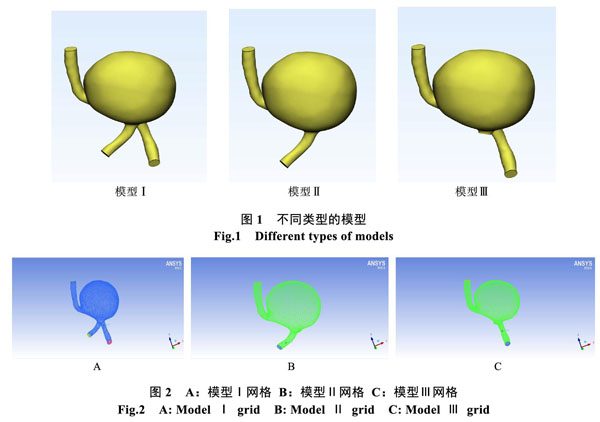
对动脉瘤模型分别进行以下处理:①阻断左侧椎动脉 ②阻断右侧椎动脉。未经过处理的动脉瘤模型为:模型Ⅰ;阻断左侧椎动脉为:模型Ⅱ;阻断右侧椎动脉为:模型Ⅲ(图1)。
1.3.2 ?网格划分
在Ansys ICEM CFD中对动脉瘤进行网格划分(图2),网格采用非结构化四面体。为保证精度,边界层设置为5层。
1.4 ?边界条件设置和数值模拟
将血流设定为牛顿流体、层流,设置血液密度为1056 kg/m3,粘度为0.0035 Pa·s[7]。设定动脉瘤壁为刚性,血液和血管壁面无滑动及渗透。椎动脉为入口,基底动脉为出口。入口速度设定为0.85 m/s,出口处的压力设定为0[8]。使用Ansys Fluent进行计算,时间步长为0.01 s,总共计算200步。
1.5 ?統计学方法
应用SPSS统计学软件。对结果进行方差分析,使用LSD-t检验对均数进行两两比较。P<0.05为差异具有统计学意义。
2 ?结果及统计学分析
2.1 ?动脉瘤内血流流线图分析
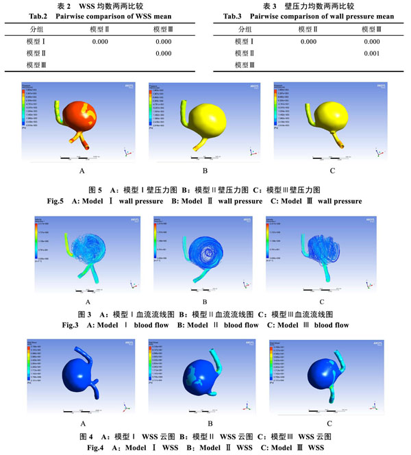
图3A中的血流从两侧椎动脉注入动脉瘤,血流注入后流场变得复杂,可见内部血流形成多个涡流,流动轨迹杂乱,最后从基底动脉流出,血流转折处流速明显上升。图3B中的血流由右侧椎动脉进入动脉瘤,血流进入动脉瘤后,多数呈层流,动脉瘤中心位置血流明显低于图3A。图3C中的血流由左侧椎动脉进入动脉瘤,血流流动与图3B相似,动脉瘤中心位置血流最低。三种模型的血流速度两两之间均有显著性差异(P<0.05,表1)。
2.2 ?动脉瘤壁切应力(Wall Shear Stress,WSS)云图分析
图4A中整个动脉瘤的WSS均处于较低的状态。在图4B中动脉瘤入口处、出口处周围以及瘤囊接近瘤顶部的位置的WSS比模型Ⅰ高。在图4C中,入口处、出口处周围以及瘤囊的一部分的WSS比模型Ⅰ高。图4B和4C的WSS变化的不同是由于两个不同的血流入射方向导致。图4B和4C中均可见到在出口血管转折处的WSS较模型Ⅰ变化最大。三种模型的WSS两两之间均有显著性差异(P<0.05,表2)。
2.3 ?动脉瘤壁压力(Wall Pressure, P)图分析
图5A中动脉瘤壁压力整体较高,在入口处、出口处周围和靠近瘤顶部的位置相对较低。图5B和图5C的动脉瘤壁压力明显低于模型Ⅰ,在图5B中,瘤囊有一小部分区域的壁压力高于其他部位,出口处周围的压力高于瘤囊。与图5B不同的是,图5C的入口处、出口处周围的压力要高,而动脉瘤囊的压力整体较低。三种模型的壁压力两两之间均有显著性差异(P<0.05,表3)。
3 ?讨论
本文通过使用数值模拟的方法,对颅内动脉瘤的CTA数据进行三维重建,并进行血流动力学分析,模拟了一例颅内动脉瘤阻断前和阻断后的血流动力学变化。本研究对阻断前后的动脉瘤内血流动力学的变化表明:阻断后,动脉瘤内血流速度和壁压力下降,WSS升高。WSS是颅内动脉瘤起始,生长和破裂过程中的关键血流动力学因素[9]。WSS近年来一直都是研究的热点,Xiang等人发现具有复杂流动模式伴有多个漩涡的动脉瘤较单个涡流的简单流动模式的动脉瘤更容易破裂,由图3可以看出,在阻断之后动脉瘤的血流模式变为简单的流动模式。WSS是血流与血管内皮间的摩擦力,其与血液特性,血流速度和血管形态由密切关系,改变血流速度,WSS会发生相应的变化,因此改变动脉瘤内和局部载瘤动脉瘤的血流动力学因素对动脉瘤的治疗有非常大的帮助。此外还发现与未破裂动脉瘤相比,破裂动脉瘤具有更大的WSS幅度和更大的低WSS区域[10]。Jou和Boussel等人实验研究表明在动脉瘤生长区域内异常低的WSS区域[11,12]。Valencia等人的报告中指出破裂动脉瘤的低WSS面积平均大于未破裂动脉瘤[13]。Fukazawa等人和Omodaka等人采用CFD方法研究了18例大脑中动脉动脉瘤术中确定破裂点,这两项研究结果获得了类似的结果,即破裂点的时间平均WSS显著低于没有破裂点的动脉瘤壁处的时间平均WSS[14,15]。Shojima等人使用CTA数据对动脉瘤进行研究,低WSS可促进生长并引发动脉瘤破裂[16]。Jou等人同样提出低WSS区域与破裂有关[17]。低WSS可以促进巨噬细胞相关的慢性炎症和动脉粥样硬化改变,由巨噬细胞造成的动脉粥样硬化炎症改变和金属蛋白酶的产生可使动脉瘤壁易于变薄并进一步破裂[18]。破裂动脉瘤破裂区域的舒张末期WSS较低,收缩期峰值压力较高,其可能的机制是低WSS导致动脉瘤壁变形和变薄,收缩期峰值的高压导致变薄壁破 ?裂[19]。低WSS导致动脉瘤壁发生变化、变薄,进而导致动脉瘤壁的破裂,在阻断之后动脉瘤的WSS升高,壁压力降低,可以避免其继续生长及破裂。
综上所述,通过椎动脉阻断的方式,基底动脉瘤的血流动力学发生显著的改变:血流速度下降、WSS升高、壁压力下降,计算结果可对临床手术规划提供借鉴。
参考文献
[1] Bederson JB I, Awad A, Wiebers DO, Piepgras D, Haley EC Jr, Brott T, Hademenos G, Chyatte D, Rosenwasser R, Caroselli C. Recommendations for the management of patients with unruptured intracranial aneurysms: a statement for healthcare professionals from the stroke council of the american heart association. Stroke. 2000; 31: 2742–2750.
[2] Bockman MD, Kansagra AP, Shadden SC, Wong EC, Marsden AL. Fluid mechanics of mixing in the vertebrobasilar system: comparison of simulation and mri. Cadiovasc Eng Technol. 2012; 3: 450–461.
[3] Boussel L, Rayz V, McCulloch C, Martin A, Acevedo-Bolton G, Lawton M, Higashida R, Smith WS, Young WL, Saloner D. Aneurysm growth occurs at region of low wall shear stress: patient-specific correlation of hemodynamics and growth in a longitudinal study. Stroke. 2008; 39: 2997–3002.
[4] Castro MA, Putman CM, Cebral JR. Computational fluid dynamics modeling of intracranial aneurysms: effects of parent artery segmentation on intra-aneurysmal hemodyna?mics. Am J Neuroradiol. 2006; 27: 1703–1709.
[5] Cebral JR, Castro MA, Appanaboyina S, Putman CM, Millan D, Frangi AF. Efficient pipeline for image-based patient- specific analysis of cerebral aneurysm hemodynamics: technique and sensitivity. IEEE Trans Med Imaging. 2005; 24: 457–467.
[6] Cebral JR, Hendrickson S, Putman CM. Hemodynamics in a lethal basilar artery aneurysm just before its rupture. AJNR Am J Neuroradiol. 2009; 30: 95–98.
[7] Paliwal, N, et al. Association between hemodynamic modi?fications and clinical outcome of intracranial aneurysms treated using flow diverters. Proc SPIE Int Soc Opt Eng, 2017; 1(2): 13-15.
[8] Yazici, B, B. Erdogmus and A. Tugay. Cerebral blood flow measurements of the extracranial carotid and vertebral arteries with Doppler ultrasonography in healthy adults. Diagnostic and Interventional Radiology, 2005. 11(4): 195.
[9] Wang, C, et al. Hemodynamic alterations after stent implantation in 15 cases of intracranial aneurysm. Acta Neurochirurgica, 2016. 158(4): 811-819.
[10] Xiang J, Natarajan SK, Tremmel M, et al. Hemodynamic- morphologic discriminants for intracranial aneurysm rupture. Stroke. 2010; 42: 144-152.
[11] Boussel L, Rayz V, McCulloch C, Martin A, Acevedo-Bolton G, Lawton M, Higashida R, Smith WS, Young WL, Saloner D. Aneurysm growth occurs at region of low wall shear stress: patientspecific correlation of hemodynamics and growth in a longitudinal study. Stroke. 2008; 39(11): 2997-3002.
[12] Jou LD, Wong G, Dispensa B, Lawton MT, Higashida RT, Young WL, Saloner D. Correlation between lumenal geometry changes and hemodynamics in fusiform intracranial aneurysms. AJNR Am J Neuroradiol. 2005; 26(9): 2357-63.
[13] Valencia A, Morales H, Rivera R, Bravo E, Galvez M. Blood flow dynamics in patient-specific cerebral aneurysm models: the relationship between wall shear stress and aneurysm area index. Med Eng Phys. 2008; 30: 329-340.
[14] Fukazawa K, Ishida F, Umeda Y, Miura Y, Shimosaka S, Matsushima S, Taki W, Suzuki H. Using computational fluid dynamics analysis to characterize local hemodynamic features of middle cerebral artery aneurysm rupture points. World Neurosurg. 2015;83: 80–6.
[15] Omodaka S, Sugiyama S, Inoue T, Funamoto K, Fujimura M, Shimizu H, Hayase T, Takahashi A, Tominaga T. Local hemodynamics at the rupture point of cerebral aneurysms determined by computational fluid dynamics analysis. Cerebrovasc Dis. 2012;34: 121–9.
[16] Shojima M, Oshima M, Takagi K, et al. Magnitude and role of wall shear stress on cerebral aneurysm: computational fluid dynamic study of 20 middle cerebral artery aneurysms. Stroke. 2004; 35: 2500-2505.
[17] Jou LD, Lee DH, Morsi H, Mawad ME. Wall shear stress on ruptured and unruptured intracranial aneurysms at the internal carotid artery. AJNR Am J Neuroradiol. 2008; 29: 1761-1767.
[18] Nixon, A. M, Gunel, M. & Sumpio, B. E. The critical role of hemodynamics in the development of cerebral vascular disease. J Neurosurg. 2010; 1(12): 1240–1253.
[19] Kono, K, et al. Hemodynamic characteristics at the rupture site of cerebral aneurysms: a case study. Neurosurgery. 2012. 71(6): 1202-8.
- 建筑工程项目管理中的成本控制研究
- 建筑工程施工质量管理问题及对策研究
- 浅析道路桥梁施工中的质量管理措施
- 新时期的小型水利工程建设质量管理工作研究
- 公路工程施工质量管理的重要性与价值
- 市政给排水管道施工中质量控制措施探讨
- 质量检查及处理措施在建筑工程中混凝土工程的作用
- 强化建筑工程监理 确保建筑工程质量
- 路桥工程施工技术及质量管理
- 建筑给排水施工中的质量通病及防治措施研究
- 浅谈干挂石材幕墙的施工管理与质量控制
- 对路桥施工技术和质量的控制措施的研究
- 模板工程的施工技术与质量控制分析
- 市政工程质量通病的成因与防治措施
- 浅谈监理工程师如何搞好工序的质量控制
- 水葱在库尔勒地区的栽培管理
- 浅论施工中如何搞好土建与安装的配合
- 上虞区水利标准化管理工作探析
- 路桥工程的施工监理分析
- 伊河东湖橡胶坝上游防渗PPP项目
- 浅析岩马水库安全度汛方案
- 以“互联网+”思维对上虞水利工程管理的思考
- 浅析建筑工程施工进度管理需要注意的问题
- 火电厂建筑工程技术管理分析
- 公路工程的现场管理工作分析
- slip out of sth
- slippage
- slippages
- slipped
- slipped disc
- slipper
- slipperier
- slipperiest
- slipperily
- slipperiness
- slipperinesses
- slippering
- slipperlike
- slippers
- slippery
- slipperyslope_
- slipping
- slippingly
- slip road
- slip roads
- slips
- slipshod
- slipshoddiness
- slipshoddinesses
- slipshodness
- 轰动震荡
- 轰去了三魂七魄
- 轰发
- 轰响
- 轰堂
- 轰堂大笑
- 轰大炮
- 轰天动地
- 轰天炮
- 轰天烈地
- 轰天裂地
- 轰天震地
- 轰子
- 轰应
- 轰打
- 轰抢
- 轰旋
- 轰炸
- 轰炸得非常厉害
- 轰炸机
- 轰烈
- 轰然
- 轰然大笑
- 轰的一声
- 轰笑